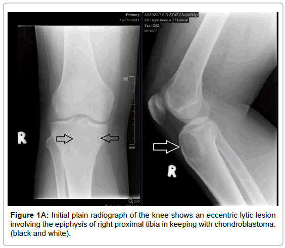
Knee Apl X Ray - Knee Horizontal Beam Lateral View Radiology Reference Article Radiopaedia Org : My 11y/o has complained of a dull, ache knee pain for awhile but after lots of playing over a week ago (no obvious injury noted) he has been in severe pain, right at the lower knee cap, radiating to the inside with significant edema.
Knee Apl X Ray - Knee Horizontal Beam Lateral View Radiology Reference Article Radiopaedia Org : My 11y/o has complained of a dull, ache knee pain for awhile but after lots of playing over a week ago (no obvious injury noted) he has been in severe pain, right at the lower knee cap, radiating to the inside with significant edema.. Ant fractures, lesions, or bony changes related to degenerative joint disease involving the distal femur, proximal tibia and fibula, patella, and knee joint may be visualized in the ap projection. It can also detect loose pieces of bone, which can cause pain. See also knee radiograph (an approach). Central ray 5 to 7 degrees cephalad at knee joint 1 inch (2.5 cm) distal to medial epicondyle. Dear tessa, lots of information here.
See also knee radiograph (an approach). The knee series is a set of radiographs taken to investigate knee joint pathology, often in the context of trauma. For most adults, the leg must be placed diagonally (corner to corner) on one 35 x 43 cm (14 x 17 inches) ir to ensure that both joints are included. It is no better and worsening. The femur is the thigh bone.

Click on the tags below to find other quizzes on the same subject.
The femur is the thigh bone. It can also detect loose pieces of bone, which can cause pain. Went home, rest, ibuprofen etc. It is no better and worsening. Normal ap and lateral knee xrays. This allows effusions to be visualised in the suprapatellar pouch. For most adults, the leg must be placed diagonally (corner to corner) on one 35 x 43 cm (14 x 17 inches) ir to ensure that both joints are included. Dr, s, venkat raman 4 years ago. Ant fractures, lesions, or bony changes related to degenerative joint disease involving the distal femur, proximal tibia and fibula, patella, and knee joint may be visualized in the ap projection. Ensure that both ankle and knee joints are 1 to 2 inches (3 to 5 cm) from ends of ir (so that divergent rays will not project either joint off the ir). Ravi kumar gupta md (radio diagnosis) from delhi university with a vision to serve the people of delhi ncr by providing latest medical investigation which are reliable , accurate & trustworthy. To correct this, externally rotate the knee. In the prone position the ir is placed under the knee and the knee is flexed 115 degrees from the horizontal axis.
For most adults, the leg must be placed diagonally (corner to corner) on one 35 x 43 cm (14 x 17 inches) ir to ensure that both joints are included. See also knee radiograph (an approach). It can also detect loose pieces of bone, which can cause pain. There are two bones below the joint line. Ant fractures, lesions, or bony changes related to degenerative joint disease involving the distal femur, proximal tibia and fibula, patella, and knee joint may be visualized in the ap projection.

My 11y/o has complained of a dull, ache knee pain for awhile but after lots of playing over a week ago (no obvious injury noted) he has been in severe pain, right at the lower knee cap, radiating to the inside with significant edema.
Slight angulation of cr will prevent joint space from being obscured by magnified image of medial femoral condyle. For most adults, the leg must be placed diagonally (corner to corner) on one 35 x 43 cm (14 x 17 inches) ir to ensure that both joints are included. Went home, rest, ibuprofen etc. Dear tessa, lots of information here. Position of patient supine or prone (prone is preferred because the knee can usually be flexed to a greater degree and immobilization is easier). This image shows parts of the bones of the knee, including the femur (the bone above the. This image shows parts of the bones of the knee, including the femur (the bone above the. Ant fractures, lesions, or bony changes related to degenerative joint disease involving the distal femur, proximal tibia and fibula, patella, and knee joint may be visualized in the ap projection. They are used primarily to confirm/exclude a fracture, or to assess the level of osteoarthritis in the knee joints (= gonarthrosis). The subjects were 257 women aged from 47 to 88 years who were outpatients at an orthopedic clinic. To correct this, externally rotate the knee. The knee series is a set of radiographs taken to investigate knee joint pathology, often in the context of trauma. Central ray 5 to 7 degrees cephalad at knee joint 1 inch (2.5 cm) distal to medial epicondyle.
My 11y/o has complained of a dull, ache knee pain for awhile but after lots of playing over a week ago (no obvious injury noted) he has been in severe pain, right at the lower knee cap, radiating to the inside with significant edema. Dear tessa, lots of information here. 2 public playlist includes this case. Normal ap and lateral knee xrays. This image shows parts of the bones of the knee, including the femur (the bone above the.

Dear tessa, lots of information here.
Position of patient supine or prone (prone is preferred because the knee can usually be flexed to a greater degree and immobilization is easier). Slight angulation of cr will prevent joint space from being obscured by magnified image of medial femoral condyle. Click on the tags below to find other quizzes on the same subject. They are used primarily to confirm/exclude a fracture, or to assess the level of osteoarthritis in the knee joints (= gonarthrosis). For most adults, the leg must be placed diagonally (corner to corner) on one 35 x 43 cm (14 x 17 inches) ir to ensure that both joints are included. In the prone position the ir is placed under the knee and the knee is flexed 115 degrees from the horizontal axis. Saral was set up in 1984 under professional guidance of dr. The subjects were 257 women aged from 47 to 88 years who were outpatients at an orthopedic clinic. The knee series is a set of radiographs taken to investigate knee joint pathology, often in the context of trauma. See also knee radiograph (an approach). This allows effusions to be visualised in the suprapatellar pouch. To correct this, externally rotate the knee. Went home, rest, ibuprofen etc.

Komentar
Posting Komentar